Fiber-Optic Sensor Advances Brain Cancer Treatment
S. Himmelstein | November 26, 2017Microbeam radiation therapy (MRT) is emerging as a technology that will enable more accurate treatment of cancers, such as brain tumors, to ensure the cancerous tissue is destroyed without damaging healthy tissue. The method uses parallel beams of X-rays, each smaller in diameter than a human hair, to cover the tumor in a precise pattern. The result is more precise targeting of doses and direction of a higher dose to the tumor itself.
Commercialization of MRT is constrained by the need for quality assurance tools to accurately detect radiation 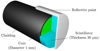 Diagram of the fiber-optic sensor. Source: University of Wollongongdosage and ensure patient safety. University of Wollongong, Australia, engineers developed a solution in the form of a new photonic detection device, just 50 microns thick, capable of measuring the dosage emitted by each of the micron-thin beams. Plastic and optic fibers were combined in the design of the photonic scintillator. The dosimeters use plastic scintillators as the radiation conversion material and plastic optical fibers as the transmission media.
“The thickness of the scintillator, or detector, need to be reduced to less than the single beam size, which would significantly reduce the number of photons generated by the scintillator and hence make the photo-detection an extremely challenging task,” said Dr. Enbang Li, from the university’s school of physics.
“The results we have achieved so far demonstrate significant steps towards the development of optical dosimeters with the potential to be applied in quality assurance of microbeam radiation therapy, which is vital if clinical trials are to be performed on human patients,” Dr. Li said. “The preclinical trials of MRT have shown that normal tissue can tolerate peak doses 100 times greater than the doses used in conventional radiotherapy.”
The next step is fabrication of fiber-optic dosimeters with 10-micron spatial resolution.