The Safety of X-Rays During Pregnancy
S. Himmelstein | July 10, 2017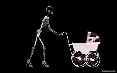 Image credit: Dream-Wallpaper.comWhat would you do if you or a loved one needed an x-ray while pregnant? Would getting one be putting the baby at risk?
No. Having an X-ray during pregnancy is generally considered safe, according to the Mayo Clinic and the American College of Radiology. It's highly unlikely that a diagnostic X-ray during pregnancy will harm a developing baby. Most X-ray exams won’t expose reproductive organs to radiation, and a leaded apron and collar can be worn to block any scattered radiation.
What Exactly Are X-Rays?
X-rays are a form of electromagnetic radiation, as are radio waves, infrared radiation, visible light, ultraviolet radiation and microwaves. They are used as a noninvasive and painless method for diagnosing disease and monitoring therapy, and supporting medical and surgical treatment planning.
The beams pass through the body and are absorbed in different amounts depending on the density of the material encountered. Dense materials, such as bone and metal, show up as white on X-rays, while the air in lungs shows up as black and fat and muscle appear as shades of gray.
X-rays are produced when electrons strike a metal target. The electrons are liberated from the heated filament and accelerated by a high voltage toward the metal target. When the electrons collide with the atoms and nuclei of the metal target, X-rays are produced.
The advent of more accurate focusing systems and more sensitive detection methods, such as improved photographic films and electronic imaging sensors, have made it possible to distinguish increasingly fine detail and subtle differences in tissue density, while using much lower exposure levels. Additionally, computed tomography (CT) combines multiple X-ray images into a 3-D model of a region of interest. The U.S. Food and Drug Administration states that X-ray imaging exams are recognized as a valuable medical tool for a wide variety of examinations and procedures.
So Is There a Problem?
According to the American College of Radiology, no single diagnostic X-ray has a radiation dose significant enough to cause adverse effects in a developing embryo or fetus. The exception is abdominal X-rays, which expose the abdomen—and baby—to radiation. High doses of radiation may cause changes in a baby's rapidly growing cells. However, the typical dose of radiation associated with a diagnostic X-ray doesn't pose this risk.
Nearly all abdominal radiographic procedures can be modified to reduce radiation exposure to a pregnant patient and her fetus, including reducing the number of images or limiting CT phases through the abdomen/pelvis, such as to a single phase. When possible, imaging should be confined to the area of interest to avoid unnecessary uterine exposure.
Improvements in imaging equipment can also contribute to reducing maternal and fetal radiation exposure. Automatic exposure control software on current generation multirow-detector CT (MDCT) scanners limits patient exposure, especially in smaller patients, by instantaneously modifying X-ray tube output to produce diagnostic images at a preset noise level. In the MDCT assessment of abdominal/pelvic trauma, the data from a single phase through the patient’s body now can provide both a comprehensive evaluation of the abdominal contents and diagnostic quality reconstructed images of the spine, eliminating the need for a second series of additional images to examine the spine. CT scanners with iterative reconstruction techniques offer potential for improving image quality and reducing radiation doses in CT relative to the currently used filtered back-projection techniques.
The average radiation dose from an abdominal X-ray is 0.7 milliGray (mGy), that from an abdominal CT scan is 8 mGy and that from a pelvic CT scan is 6 mGy. A dose of 20 mGy represents an additional projected lifetime risk of about 40 additional cancers or less per 5,000 babies, or about 0.8 percent. In other words, there is an over 99 percent likelihood the fetus will be unaffected by the radiation.