Vector flow imaging aids diagnostics for pediatric heart patients
S. Himmelstein | April 11, 2019Detection of congenital heart defects in infants typically requires ultrasound techniques to provide critical data on cardiac valve function. The technology has its limitations, as ultrasound cannot accurately obtain details of blood flow within the heart, due to the inability to align the ultrasound beam with blood-flow direction. Vector flow imaging (VFI), an ultrasound-based imaging method, was recently demonstrated to visualize the direction 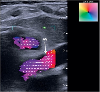 Transthoracic vector flow imaging in Patient 1 demonstrates a healthy anatomy with laminar flow across the aortic valve (AV). No turbulence is observed. Source: R. Thomas Collins II et al.and speed of blood flow, independent of the orientation of the ultrasound probe.
Researchers from Cincinnati Children's Hospital Medical Center, Stanford University, University of Arkansas and Arkansas Children's Research Institute applied VFI for the first time to pediatric patients to generate detailed images of the internal structure and blood flow in hearts. The images can be still or moving and can be captured from any angle. The demonstration used the bk5000 ultrasound system from BK Medical, which has been advancing VFI technology.
Tests were successfully performed on two pigs, one with normal cardiac anatomy and one with congenital heart disease due to a narrow pulmonary valve and a hole within the heart. The vector flow images were then compared to direct examination of the pigs’ hearts. The researchers next used the imaging system to take cardiac images of two three-month-old babies, one with a healthy, structurally normal heart and one with congenital heart disease marked by an abnormally narrow aorta. With both patients, the technology-enabled total transthoracic imaging of tissue and blood flow at a depth of 6.5 cm, and abnormal flow and detailed cardiac anomalies were clearly observed in the patient with congenital heart disease.
The study published in Progress in Pediatric Cardiology confirms that the commercially available technology can function as a bedside imaging method to assess flow characteristics in both healthy and diseased hearts in a broad range of infants and children.