Watch: Microfluidic Bleeding Platform for Wound Healing Research
S. Himmelstein | February 08, 2018A miniature self-sealing model system for studying the bleeding and clotting of wounds is considered the 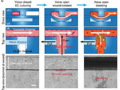 Schematic of the microfluidic system. Source: Nature Communicationsfirst to reproduce all facets of blood vessel injury occurring in the microvasculature. The system, which simulates blood loss due to trauma, clot formation by whole blood and repair of the blood vessel lining, could find application as a drug discovery platform and potential diagnostic tool.
The polydimethylsiloxane-based microfluidic bleeding time device consists of a layer of human endothelial cells cultured on top of a pneumatic valve. The "wound," measuring 130 micrometers across, is created by activating the valve, allowing donated human blood to flow through.
As seen in the video above, most of the blood cells are grey: erythrocytes are grey donuts, while platelets are smaller specks. Red-stained cells are actually white blood cells, and a green extracellular "glue" evident at the top of the wound is fibrin, which holds the clot together.
In real time, it takes about eight minutes for blood flow into the wound to stop. Without the endothelial cells, the blood flow does not stop.
The system responds to manipulation by drugs and other alterations that reproduce clotting disorders. Blood from hemophilia A patients forms abnormal clots and shows extended bleeding time in the model.
Scientists from Georgia Institute of Technology, Emory University, Blood Center of Wisconsin, VA Boston Healthcare System and Harvard Medical School participated in this research, which is published in the journal Nature Communications.