Modern approaches to bioprinting technology
Jody Dascalu | November 18, 2025Bioprinting combines principles from biology, materials science and manufacturing to create living structures from cells. Instead of printing with plastic or metal, these systems use bioinks made from cells and hydrogels to build tissue-like materials layer by layer. The technology is already reshaping regenerative medicine, tissue engineering and pharmaceutical research. Printed cartilage patches can restore damaged joints, and miniature liver models are being used to study drug toxicity without animal testing. The central challenge is organizing delicate cells into stable, functional architectures that remain viable and behave like natural tissue. Progress in printer design, bioink chemistry and process control continues to push the field closer to reliable, clinically relevant applications.
Core principles of bioprinting materials
Bioprinting relies on bioinks, which are mixtures of living cells and hydrogels that provide a temporary structural environment. The physical properties of these materials are critical. The ink must be viscous enough to hold its shape after printing but fluid enough to pass through the nozzle without damaging the cells. High pressure or shear stress can reduce cell viability, while poor mechanical strength can cause printed layers to collapse. After deposition, the material needs to allow nutrient and oxygen diffusion to support continued growth. Bioprinting approaches generally fall into two categories: scaffold-based methods that use supportive matrices for structure, and scaffold-free methods that depend on the natural aggregation and fusion of cells. These parameters define print quality, biological function and long-term stability.
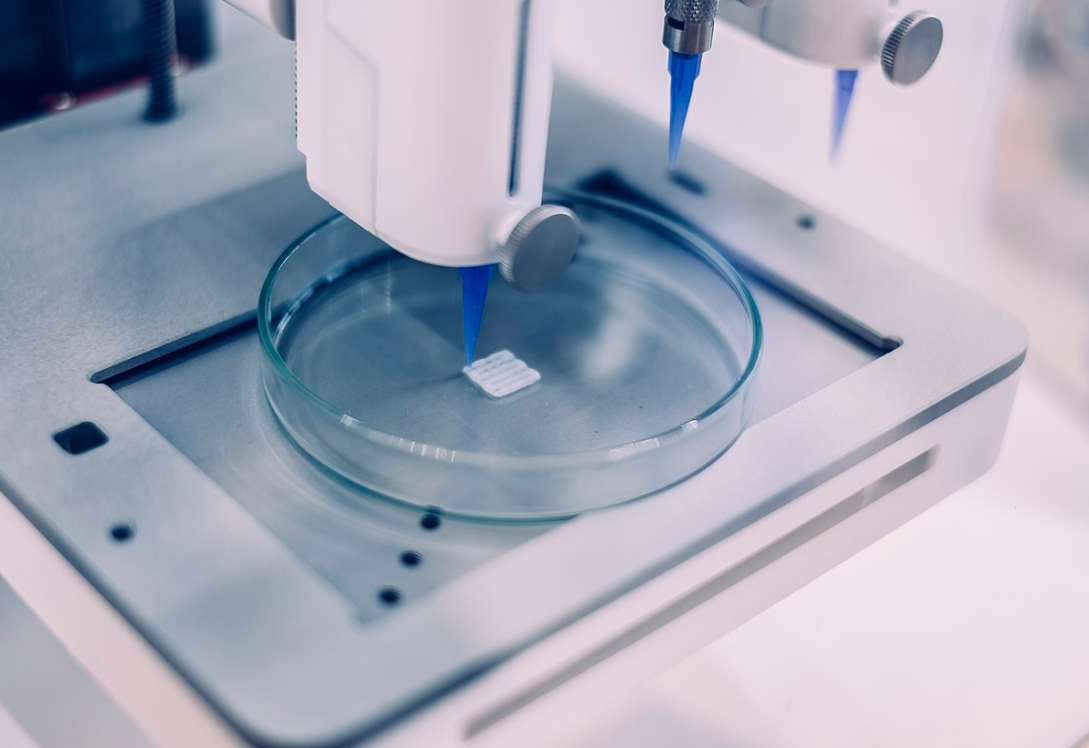 Bioprinting a square of organism in a petri dish. Source: Philip Ezze/CC BY-SA 4.0
Integrating biology with manufacturing
Inkjet bioprinting
Inkjet bioprinting is one of the earliest and most widely studied methods in the field. It operates on the same basic principle as a conventional inkjet printer, ejecting small droplets of bioink through a nozzle onto a substrate in a controlled pattern. The actuation mechanism can be either thermal or piezoelectric. Thermal systems briefly heat the bioink to create vapor bubbles that force droplets out of the nozzle, while piezoelectric systems use mechanical pressure from a crystal actuator.
This technique works best with low-viscosity bioinks that flow easily and do not contain large cell aggregates or particles that could block the nozzle. Its main advantages are high precision, relatively low cost and the ability to deposit multiple cell types in defined patterns. Because each droplet can be placed with micrometer-scale accuracy, inkjet systems are well suited for creating thin, layered tissues or for testing cell behavior in structured microenvironments.
However, the simplicity of the system comes with trade-offs. The limited viscosity range restricts the types of materials that can be printed, and nozzle clogging remains a frequent technical challenge. In thermal inkjet systems, the transient heating step can also stress or kill a portion of the cells, although modern designs minimize this risk by using very short heating pulses. The droplet-based nature of the method also makes it difficult to build thick, mechanically stable constructs without additional crosslinking or structural support.
Despite these limitations, inkjet bioprinting remains valuable for applications that require fine control and moderate throughput. It has been used to fabricate skin tissue models, vascular patterns and small organoid arrays for drug testing. Because it combines precision with scalability, it continues to serve as a key platform for high-throughput biological research and for exploring how printed microarchitectures influence cell growth and differentiation.
Extrusion bioprinting
Extrusion bioprinting is currently the most common and versatile method in tissue fabrication. Instead of ejecting droplets, it continuously deposits a filament of bioink through a nozzle using pneumatic pressure or a mechanical system such as a piston or screw drive. The extrusion process allows for a wider range of bioink viscosities and higher cell densities than inkjet systems, which makes it suitable for building thicker, more robust structures.
This technique prioritizes material flexibility and scalability over resolution. The printed strands are generally larger, which limits fine detail but allows the construct to maintain mechanical integrity. The continuous flow also introduces shear stress that can affect cell viability, so parameters such as pressure, nozzle diameter and speed must be carefully balanced. Despite these challenges, extrusion systems can print multiple materials simultaneously, integrate temperature control and support on-the-fly crosslinking to stabilize each layer as it is deposited.
Because of its reliability and adaptability, extrusion bioprinting has become the workhorse of the field. It is commonly used to produce cartilage, bone and vascular grafts, as well as larger composite tissues that require structural stability. Advances in multi-head printers and bioink formulations continue to expand its potential for clinical applications.
Laser-assisted bioprinting
Laser-assisted bioprinting uses focused light energy to transfer precise volumes of bioink onto a receiving substrate. The system typically includes a laser source, a transparent donor slide coated with a thin layer of bioink, and a collector plate positioned below. When the laser pulse strikes the donor layer, it generates a small pressure bubble that propels a microdroplet of material onto the target surface.
Because there is no nozzle, the technique avoids clogging and can handle a wide range of viscosities. It also achieves extremely high spatial resolution, often in the range of tens of micrometers, making it suitable for applications that demand fine control over cell placement. Cell viability is typically high, as the short and localized laser energy minimizes thermal damage.
The main drawbacks are cost, complexity and limited printing speed. Calibration must be precise, and throughput is significantly lower than in extrusion or inkjet systems. However, for specific uses such as neural tissue engineering, vascular network patterning and research on cell-to-cell interactions, laser-assisted bioprinting provides a level of accuracy that other methods cannot match.
Emerging strategies and ongoing challenges
Recent advances in bioprinting focus on overcoming the structural and biological limitations of current techniques. Hybrid systems combine multiple printing modes, such as coaxial extrusion for producing hollow fibers that mimic blood vessels, or embedded methods like FRESH printing, which use a temporary support bath to build freeform, self-supporting tissues. These approaches allow for more complex geometries and improve nutrient delivery in larger constructs.
Control systems are also becoming smarter. Machine learning models can optimize print paths, pressure, and temperature in real time to reduce stress on cells and improve consistency. Imaging feedback systems now track layer formation as printing occurs, enabling automatic corrections during fabrication.
Despite these advances, key challenges remain. Achieving full vascularization in thick tissues is still a major obstacle, as is standardizing bioink properties across laboratories. Scaling from small tissue samples to functional organs also requires new strategies for nutrient transport, structural integration and long-term viability.
Future outlook and medical potential
Bioprinting is moving steadily from experimental prototypes toward practical medical applications. Each printing technique presents trade-offs among resolution, speed and cell survival, but together they are shaping a new manufacturing paradigm for living materials. As the technology matures, advances in 4D printing, adaptive materials and personalized biofabrication may bring the long-term goal of functional, patient-specific organs within reach.