MRI: Led by Physics, Followed by Lawsuits
Bill Schweber | February 25, 2016Magnetic resonance imaging (MRI) is a powerful technique for non-invasively creating detailed images of the inside of a body, and with relatively little risk. The principles of MRI are based on atomic-level subtleties and not at all obvious or easy to explain. By contrast, two other common imaging technologies—X-rays and ultrasound—are a bit more straightforward: X-rays were discovered by accident and ultrasound is an implementation of well-understood echo principles. MRI, however, came about only after some deep discoveries in quantum physics which then were extended by researchers who imagined how to adapt and build on those principles.
MRI was originally called nuclear magnetic resonance or NMR imaging. The "nuclear" part was dropped because it scared people, who thought they were being irradiated, which is not the case at all. MRI uses magnetic fields and non-ionizing radio frequency waves at very low levels, and has no relation to the public's concept of "nuclear."
A Moment and a Magnet
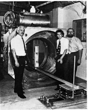 The first human MRI scanner machine, constructed in 1977. Left to right are doctors Damadian, M. Goldsmith and L. Minkoff. Image source: Fonar Corp. Two key concepts are necessary to understand the MRI concept. First, there's the atomic basis, the magnetic moments of an atom's nucleus tends to align either parallel or anti-parallel to an external magnetic field. Like a common spinning top, it precesses about the direction of the magnetic field alignment and at a frequency that depends on the magnetic field's strength and the atom’s nuclear magnetic moment.
The magnetic moments of nuclei can be induced to flip their orientation if they absorb energy from an imposed electromagnetic wave of the right frequency. They also emit this absorbed energy when they fall back to their lower-energy orientation, a transition that can be detected when the energy beam is exposed to a radio frequency signal as it travels through the magnetic field. Tuning either the external magnetic field or the radio frequency can produce resonance.
Second, there’s the biological basis. The concept and realization of a complete resonance-based imaging system was developed and implemented by Dr. Raymond Damadian, a physician and experimenter working at Downstate Medical Center in Brooklyn, New York. He realized that because tumors contain more water and thus more hydrogen atoms than healthy tissue, the MRI-induced radio signals from cancerous tissue will differ from those of healthy tissue. Specifically, they will have a longer relaxation time.
His first full-body MRI scan in 1977 took nearly five hours to produce an image; today's scans are nearly instantaneous due to advances primarily in the numeric-computational "horsepower." Damadian named that first MRI scanner the "Indomitable," and it is now in the Smithsonian Institution.
Not a Selfie
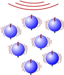 In the presence of an intense imposed magnetic field, the protons absorb energy and their axes line up to create a single magnetic vector; they radiate this energy when the magnetic field is removed.
 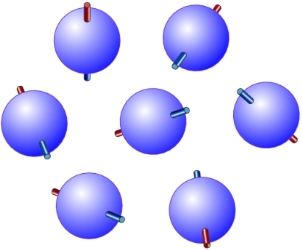 Without an external magnetic field, protons (hydrogen nuclei) are randomly oriented. Image source (1-4) : U.S. National Library of Medicine, National Institutes of Health.For imaging purposes, the hydrogen nucleus (a single proton) is used because of its abundance in water and fat. Under normal circumstances, these hydrogen proton “bar magnets” spin in the body with their axes randomly aligned. When the body is placed in the magnetic field of the MRI scanner, the axes of these protons line up (as shown in the figures). This uniform alignment creates a magnetic vector oriented along the axis of the MRI scanner.
MRI scanners use different magnetic-field strengths beginning at about 0.5 and 3 tesla (T; 1 T = 10,000 gauss) as a minimum value; higher field strengths result in improved resolution. This field intensity can be created only using high-current superconducting magnets cooled by liquid helium to nearly absolute zero.
When additional energy in the form of a radio wave is added to the applied magnetic field, the magnetic vector is deflected. The radio frequency (RF) wave that causes the hydrogen nuclei to resonate is dependent on the target element (here, hydrogen) and the strength of the magnetic field. To make the MRI work, the strength of the magnetic field in smaller, gradient magnets (about 200 gauss) is electronically altered from head to toe by using a series of gradient electric coils. By altering the local magnetic field, different slices of the body resonate as the different frequencies are applied.
In practice, the MRI scanner contains two magnets. The first one is static, and causes all of the body's water molecules to align in one direction. The second magnetic field is then switched on and off in a series of quick pulses. These pulses cause each hydrogen atom to alter its alignment and then revert to its original, relaxed state when switched off. As a result of the gradient coils being switched on and off, the magnetic coil vibrates. This creates a loud knocking sound inside the scanner.
When the external RF source is switched off, the aggregate magnetic-moment vector returns to its resting state and the proton emits an RF signal. This is the signal that creates the raw data for images. An array of sensitive receiver coils with high-gain, low-noise amplifiers is located around the body and acts as antennas to detect the emitted signal. (Note that MRI does not inherently create a two-dimensional image; instead, the intensity of the received signals is plotted on a gray scale and cross-sectional images are built up using image-processing algorithms.)
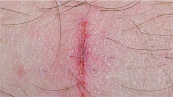
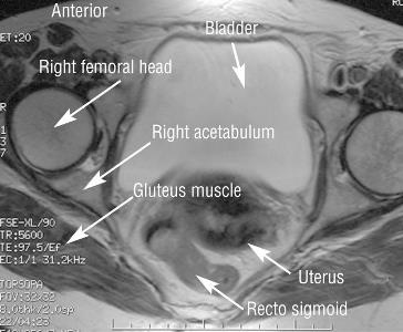 The same pelvis MRI image, but using a T2 wave.
 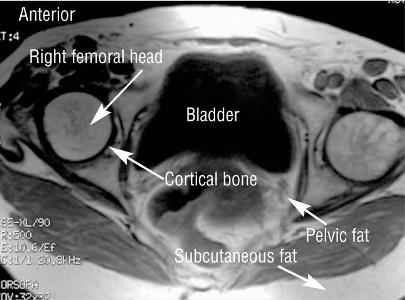 An axial image of a pelvis, using T1 MRI wave. The time taken for the protons to fully relax is measured in two ways. T1 relaxation is the time that the magnetic vector takes to return to its resting state. T2 relaxation is the time needed for the axial spin to also return to the resting state. These are shown in the accompanying images.
Revolutionizing Basic Diagnostics
Advanced MRI designs have added to capabilities of the basic concept and implementation. Because protons in different body tissues relax and return to their normal spins at different rates when the transmitted RF pulse is switched off, the scanner can be adjusted to distinguish among tissues. Additional magnetic fields can be used to localize body structures in three dimensions, and multiple radio-frequency pulses can be transmitted in sequence to highlight specific tissues or abnormalities.
Application-specific MRI variations now exist, such as diffusion MRI and functional MRI. Diffusion MRI measures how water molecules diffuse through body tissues; some diseases, such as a stroke or tumor, can restrict this diffusion, so this method is often used to diagnose them. Functional MRI (fMRI) measures changes in blood flow in different parts of the brain by assessing blood-oxygen-level dependent (BOLD) contrast. As neurons use more oxygen when they're active, these BOLD signals are an indication of brain activity, and fMRI has had a major impact on studies of the brain.
MRI is an excellent example of where developments in one or more fields (namely, superconduction and supercooling, digital computing, low-noise RF amplifiers) enable advances in unrelated fields (medical imaging). The technology also serves up an example of how a fundamental discovery with no apparent practical use can become the basis for a revolutionary new technique, product and industry.
Nobel Prizes
Obviously, no single person or team invented the MRI in isolation; it is the culmination of decades of scientific progress and understanding. MRI technology begins with the discovery of a quantum-physics phenomenon called nuclear magnetic resonance (NMR) in 1937 by Isidor I. Rabi, a Polish-born American physicist. He showed that atomic nuclei indicate their presence by absorbing or emitting radio waves when exposed to a sufficiently strong magnetic field. For this work, he received the Nobel Prize in Physics in 1944.
By 1946, Felix Bloch at Stanford University and Edward Purcell at Harvard University independently extended the NMR phenomenon beyond nuclei to properties of atoms and molecules in solids and liquids. Instead of individual atoms or molecules of Rabi’s molecular beam, this became the basis for NMR-based spectroscopy to study the composition of chemical compounds (and garnered a Nobel Prize in Physics for them in 1952).
Subsequently, Paul Lauterbur, a professor of Chemistry at the State University of New York at Stony Brook, proposed a technique and experiments which took the single dimension of NMR-based spectroscopy to two dimensional-imaging. In 1973 his work produced the first NMR image (of a test tube). Around the same time, Peter Mansfield of Nottingham, England, showed how gradients in the magnetic field could be mathematically analyzed, making a useful, fast-imaging technique possible. The two received the 2003 Nobel Prize in Physiology or Medicine for their work.
Meanwhile, in 1970, Raymond Damadian found that different kinds of animal tissue emitted signals that vary in length, and that the signals from cancerous cells last much longer than those from non-cancerous tissue. He filed for and received a patent in 1974 for an "Apparatus and Method for Detecting Cancer in Tissue," the first MRI-related patent. He completed construction of a whole-body MRI scanner in 1977. His research team built the entire machine themselves, including winding the magnet coils and plumbing the superconducting-magnet subsystem.
Success, then Controversy
The first test was a failure, but Damadian and his team realized the problem: he was physically too large for the sensor array. One of his graduate students, Larry Minkoff, volunteered to take his place, and after nearly five hours, the first human scan was complete. (Read Damadian's paper on the construction of the machine, the underlying analysis and the data.)
In 1978, Damadian formed Fonar ("Field Focused Nuclear Magnetic Resonance") and in 1980, he produced the first commercial unit. However, for technical reasons, he had to abandon his original technique in favor of a variation developed by Lauterbur and Mansfield.
That's where things became messy: Damadian and Fonar obtained royalties on their patents and settled with many large companies to use the technology. General Electric would not acknowledge the validity, however, but Fonar eventually prevailed and received a $129 million ruling against GE.
The story doesn't end there, as Lauterbur and Mansfield's Noble Prize award did not include Damadian (Nobel rules allow for up to three recipients). The dispute over "proper credit" for MRI has gone on for many years, with vocal adherents on both sides. Even the American Physical Society (APS) page on MRI (Reference 2) gives full names and details for the fundamental research but changes to an anonymous, passive tone when it gets to specifics of the first MRI. It makes no mention of Damadian; similarly, detailed and lengthy reference from The Journal of Cardiovascular Magnetic Resonance mentions him once, and then only as part of a list of contributors to MRI development.
Adherents on both sides of the controversy mounted public campaigns, with letters in scientific journals, press conferences, debates at conferences and even full-page newspaper ads (this was before the Internet). All a rather unusual spectacle for the scientific community.
References
- http://www.fonar.com/pdf/TL/doc13.pdf
- https://www.aps.org/publications/apsnews/200607/history.cfm
- http://www.scmr.org/assets/files/members/documents/JCMR/008/LCMR_i_008_04_tfja/LCMR_i_8_04_O/LCMR_A_175489_O.pdf
- http://www.two-views.com/mri-imaging/history.html#sthash.zXsFeG0s.dpbs
- http://inventors.about.com/od/mstartinventions/a/MRI.htm
- http://www.ncbi.nlm.nih.gov/pmc/articles/PMC1121941/