Mini MRI produces keener knee scans
S. Himmelstein | July 02, 2019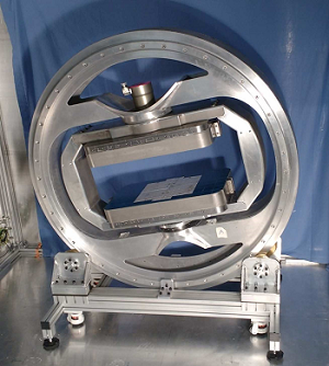 A scaled-down version of the conventional MRI scanner promises to improve the accuracy and speed of knee injury diagnosis. The compact equipment addresses two limitations encountered with current scanners: the inability to clearly image certain structures in the knee, and long wait times for diagnostic tests, a problem for patients in the U.K.
Hospital-based MRI systems do not produce sharp pictures of tendons, ligaments and meniscus. The brightness of these structures in images is governed by the angle between collagen fibers and the magnetic field of the scanner. Imperial College London researchers revised the orientation of the magnet and subsequently that of the magnetic field in the mini MRI scanner with the aid of motors and sensors, enabling observation of the pattern and condition of collagen in knee structures as a prerequisite for prescribing treatment.
The mini MRI accurately detected ligament and tendon damage in animal models. After successful trials on humans, the device could help diagnose conditions such as anterior cruciate ligament injuries, and its small size means it could eventually be used in local clinics.