CT data library compiled to support reduced radiation dose research
S. Himmelstein | April 28, 2020Computed tomography (CT) technology helps clinicians see detailed images of the internal anatomy in a process that results in accumulation of radiation doses to the patient. Mayo Clinic scientists have assembled a CT data library to help meet the goal of acquiring the required image detail but with reduced radiation doses.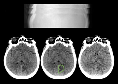 Sample X-ray projection images through a patient’s head are used to reconstruct CT images of the patient’s brain. Source: Mayo Clinic
Medical imaging researchers can use the publicly accessible library to develop, validate and optimize algorithms and enhance imaging hardware to produce peak-quality CT images using low radiation doses. The library includes reconstructed images from 300 patient exams and X-ray projection data used to create cross-sectional images, all of which are critical for the development of advanced image reconstruction techniques, including those using artificial intelligence.
The Low Dose CT Imaging and Projection Data collection, housed at The Cancer Imaging Archive hosted by the University of Arkansas for Medical Sciences, includes 100 non-contrast-enhanced head CT exams, 100 non-contrast-enhanced chest CT exams and 100 contrast-enhanced abdominal CT exams. Half of each data set is derived from a CT scanner manufactured by Siemens Healthcare and the other half from a scanner manufactured by GE Healthcare.