Microscope Add-on Enhances 3D Imaging of the Brain
S. Himmelstein | September 18, 2018Technology developed at Tel Aviv University improves 2D and 3D imaging of neuronal activity in living brains of living animals. The microscopy advance seamlessly enables integration of the fastest 3D imaging solution available to help researchers better understand brain dynamics and discover new treatments for stroke, epilepsy and other health problems. PySight uses open-source software and commercially available hardware to give laser scanning microscopes a photon counting feature.
The multiphoton microscopy laser-based technique can image deep into tissue and is often applied to study the rapid activity patterns of neurons, blood vessels and other cells at high resolution over time. The method uses laser pulses to excite fluorescent probes, eliciting the emission of photons, some of which are detected and used to form 2D and 3D images.
Efforts to capture the full breadth of neuronal activity with multiphoton microscopy force researchers to 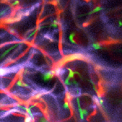 Brain vasculature in an anaesthetized mouse captured using Pysight. Source: Pablo Blinder, Tel Aviv Universityimage faster, resulting in fewer and fewer photons available to form images, so that frames become progressively dimmer and obscure details of interest. Another challenge is that implementation of photon counting requires extensive electronics knowledge and custom components, posing barriers to its adoption.
PySight overcomes these challenges by providing high spatiotemporal resolution while producing a data stream that scales with the number of detected photons, not the volume or area being imaged. The system stores the precise detection time of each photon to facilitate rapid imaging of large volumes over long sessions, without compromising spatial or temporal resolution.
Photon arrival times are generated by a multiple-event time digitizer, or multiscaler, which records the times with a precision of 100 picoseconds. An off-the-shelf resonant axial scanning lens changes the focal plane hundreds of thousands of times per second. The researchers used this setup to rapidly scan the laser beam across different depths within the brain and to reconstruct continuous 3D images.
The software is open source and provides direct access to photon arrival times, enabling other scientists to add new features and meet their specific needs.
The research is published in Optica.