Watch How 3D-printed Aortas Benefit Heart Valve Replacement
S. Himmelstein | March 08, 2018Successful prosthetic heart valve implantation requires matching of the implanted device to the patient 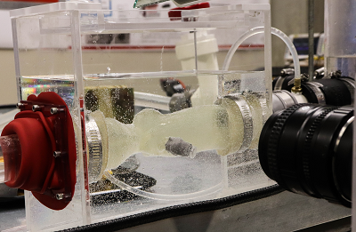 A 3D-printed replica of a patient’s aorta is tested in a high-tech heart simulator. Source: The Ohio State Universityanatomy and placing the device so as to prevent leaks and clot formation. To improve outcomes of mitral valve replacement procedures, researchers at The Ohio State University Wexner Medical Center are using CT scans to model a patient’s aorta, then 3D-print a replica based on the calcification in a patient’s tissue.
The model aortas are tested in a special simulator that replicates hemodynamic parameters such as flow, pressure and turbulence characterizing left ventricular outflow. Contraction and expansion match each patient’s anatomy as a blood-like fluid is pumped through the simulator. Clinicians and researchers can observe the optimal type of valve and specific placement for that patient in order to avoid the aforementioned complications.
Without this technology, doctors use their best judgment based on CT imaging, the patient’s history and experience with various valve options. Sometimes, potential complications surface after the aortic valve is placed.