Watch How a Map App Fills Stem Cell Research Gap
S. Himmelstein | February 16, 2018Medical researchers are culturing stem cells in pursuit of disease therapies, tissue and organ engineering and personalized medicines. A challenge to realizing these diverse goals is the lack of measurement tools for stem cell production which could help gauge which new discoveries are safe and effective.
An image analysis app released by the U.S. National Institute of Standards and Technology is designed to fill this research gap. The Web Image Processing Pipeline (WIPP) app allows a user to interact with microscopic views of macroscopic objects. With WIPP, a cell culture is divided into real locations that can be explored and referenced with a system very much like GPS points or the quadrants on a geographic map. It also allows a user to examine what’s happening in a petri dish over time, from any point of view they choose.
The system was designed for work with induced pluripotent stem (iPS) cells -- those taken from the tissue of 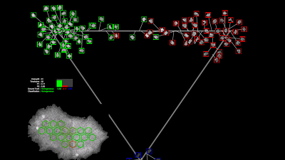 WIPP allows researchers to examine and investigate cells in many new and dynamic ways. Source: Jose Garcia/NISTan adult and processed to behave like an embryonic stem cell. iPS cells can be used to make new cells from all three of the basic human body layers: ectoderm (skin), endoderm (gastrointestinal and respiratory tracts) and mesoderm (blood and bone). iPS cell technologies are still relatively new, and it is unclear which tissue types derived from iPS cells will have the greatest impact first. Researchers are seeking ways to effectively and reliably measure progress as cells become different types during processing.
To use WIPP, researchers upload their data to a server, which carries out some computational operations, while others are carried out on a desktop with the web browser. The entire WIPP system is open access and in the public domain, although researchers’ own datasets remain private for specific projects until they choose to make them public.
Sections of the cell culture dish where a colony is being grown are photographed in sequence via microscope. The images are then stitched together to form a view of the entire dish at each point in time. Each of those complex composite, single-time-point images are then run like a movie, revealing the cells’ entire history.
WIPP incorporates the computations behind the images, pulling in the analytical data to match each view of the experiment. This allows users to collect the key measurements about shape, size, texture features, fluorescence intensities, contrast and rates of change in these measurements, and to analyze the cell population within each colony over time and space.