Camera Spots Esophageal Pre-Cancers Earlier
John Simpson | September 21, 2016Research from a team at the Cambridge Research Institute, in the UK, could lead to a new way of detecting precancerous changes that could develop into esophageal cancer.
An earlier diagnosis can mean availability of more treatment options with a greater likelihood of success. For cancers that develop through cellular changes linked to other conditions, early detection can even mean the chance to prevent a cancer from developing. But spotting the earliest signs of a disease isn’t easy.
Combining imaging, engineering and biochemistry, researchers led by Dr. Sarah Bohndiek have developed a new way to take pictures of cells using a specialized camera and fluorescent light given off by a dye—offering a glimpse at what happens when good cells go bad.
The team's study into the early detection of esophageal cancer focused on the presence of Barrett’s esophagus. The condition causes changes to the cells that line the esophagus, meaning that patients with Barrett’s have an increased risk of developing esophageal cancer. Because of this, they are often closely monitored using endoscopy, a technique that captures videos of the inside of the esophagus.
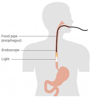 Diagram of an endoscopy procedure. Image credit: Cancer Research UK.Endoscopies use a normal white light camera to scrutinize the surface of the esophagus for the early changes that could become cancer. But, unlike many cancers, which form visible lumps, the precancerous changes caused by Barrett’s appear as flat patches, sometimes with very little change in tissue color.
To improve the visibility of the precancerous changes, the researchers first conducted tests using a fluorescent dye that sticks to specific molecules on esophageal cells. When a specific color of light was shined on the cells in the lab, the targeted dye gave off a different color of light back to the camera to indicate if the dye had stuck or not. The molecule the dye stuck to was found only on healthy esophageal cells, not the precancerous regions.
But there was a problem: the cells lining the esophagus were also fluorescent, so the difference in brightness between the different cells was very low.
To overcome this challenge, the team selected a new targeted dye that gives off a fluorescent color different to that of the esophagus. The color chosen for the dye was near-infrared—the range of light between familiar red light and invisible infrared light.
To capture images of this dye, the researchers used a specialized camera that can be turned into an endoscope to "see" the near-infrared light. The device was tested in the lab to determine if it could spot the unhealthy cells. This included pinpointing the smallest object the endoscope could detect (its resolution), the smallest amount of dye it could detect (its sensitivity) and the largest object that could be seen in a single image.
To check that the endoscope accurately picked up the dye, the researchers took images of the dye sprayed onto human esophageal tissue samples in the lab and compared them to images captured with a standard near-infrared camera. The images closely matched, indicating that the dye approach works on real cells.
After further development and safety testing of the device, the team hopes to carry out a clinical trial of this technique in people with Barrett’s esophagus.


