Supervised Autonomous Robotic Soft-Tissue Surgery Demonstrated
Engineering360 News Desk | May 17, 2016Surgeons and scientists from Sheikh Zayed Institute for Pediatric Surgical Innovation at Children's National Health System have demonstrated that supervised autonomous robotic soft-tissue surgery on a live subject (in vivo) in an open surgical setting is feasible and outperforms standard techniques in a dynamic clinical environment.
A study published in Science Translational Medicine reports the results of soft-tissue surgeries conducted on both inanimate porcine tissue and live pigs using proprietary robotic surgical technology—the Smart Tissue Autonomous Robot (STAR)—developed at Children's National. This technology removes the surgeon's hands from the procedure, instead utilizing the doctor as supervisor, with soft-tissue suturing autonomously planned and performed by the STAR robotic system.
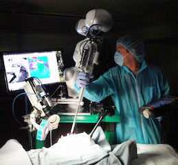 STAR employs a bedside lightweight robot arm extended with an articulated laparoscopic suturing tool for a combined eight-degrees-of-freedom robot. Image credit: Children's National Health System."Our results demonstrate the potential for autonomous robots to improve the efficacy, consistency, functional outcome and accessibility of surgical techniques," says Dr. Peter Kim, vice president and associate surgeon-in-chief. "The intent of this demonstration is not to replace surgeons, but to expand human capacity and capability through enhanced vision, dexterity and complementary machine intelligence for improved surgical outcomes."
Soft tissues are those that connect, support or surround other structures and organs of the body, such as tendons, ligaments, fascia, skin, fibrous tissues, fat, synovial membranes, muscles, nerves and blood vessels. Currently more than 44.5 million soft-tissue surgeries are performed in the U.S. each year.
While robot-assisted surgery (RAS) has increased in healthcare settings, the execution of soft-tissue surgery has remained entirely manual, largely because of the unpredictable, elastic and plastic changes in soft tissues that occur during surgery, requiring the surgeon to make constant adjustments. To overcome this challenge, STAR uses a tracking system that integrates near-infrared fluorescent markers and 3D plenoptic vision, which captures light field information to provide images of a scene in three dimensions.
This tracking is combined with another STAR innovation, an intelligent algorithm that guides the surgical plan and autonomously makes adjustments to the plan in real time as tissue moves and other changes occur. The STAR system also employs a bedside lightweight robot arm extended with an articulated laparoscopic suturing tool for a combined eight-degrees-of-freedom robot.
To compare the effectiveness of STAR to other available surgical procedures, the study included two different surgeries performed on inanimate porcine tissue (ex vivo), linear suturing and an end-to-end intestinal anastomosis, which involves connecting the tubular loops of the intestine. The results of each surgery were compared with the same surgical procedure conducted manually by an experienced surgeon and by RAS with the Da Vinci Surgical System.
Intestinal anastomosis was also performed on the living subjects (in vivo) in the study. Four anastomosis surgeries on living pigs were conducted using STAR technology, and all subjects survived with no complications. The study compared these results to the same procedure conducted manually by an experienced surgeon using standard surgical tools.
The comparisons showed that supervised autonomous robotic procedures using STAR were superior to surgery performed by experienced surgeons and RAS techniques—whether on static porcine tissues or living specimens—in areas such as consistent suture spacing, which helps to promote healing, and in withstanding higher leak pressures.
Kim says that since supervised autonomous robotic surgery for soft-tissue procedures has been proven effective, a next step in the development cycle would be further miniaturization of tools and improved sensors to allow for wider use of the STAR system. He adds that, with the right partner, some or all of the technology can be brought into the clinical space and bedside within the next two years.