New light-triggered patch offers fast, leak-proof sealing in neurosurgery
Marie Donlon | February 09, 2026Researchers from South Korea’s Pusan National University have created a light-responsive, monolithic Janus dural patch that offers leak-proof sealing in neurosurgery.
The team explained that a common neurosurgical complication dubbed durotomy occurs when there is a tear in the dura mater, which is the protective membrane surrounding the brain and spinal cord. This damage can reportedly cause cerebrospinal fluid (CSF) leakage, resulting in delayed healing, headaches and infection, thus making a watertight dural closure, such as the one from Pusa National University, essential.
 Source: Pusan National University
While tissue adhesives are currently being examined as alternatives to suturing for dural closure as they provide simpler and faster application, the majority of existing glue-based sealants suffer from excessive swelling, thereby resulting in mass effect and unwanted tissue adhesion. This, the researchers explained, can lead to postoperative complications.
Researchers have examined Janus tissue patches, which include two distinct surfaces — one surface that strongly adheres to tissue and another surface that prevents unwanted adhesion. Yet, most existing Janus patches tend to rely on several materials as well as multi-step fabrication processes, both of which limit their practical use.
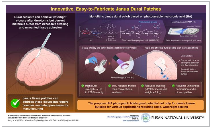
As such, the team used photocurable hyaluronic acid (HA) through a simple approach to develop their patch. “Made from natural biopolymer hyaluronic acid, our dural patch provides strong wet adhesion, along with a lubricating surface that prevents unwanted tissue adhesion, after exposure to non-toxic visible light,” the team explained.
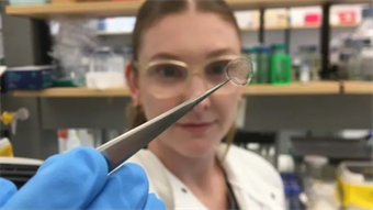
The team selected HA for its biocompatibility and natural anti-adhesive and lubricating properties, then modified it with photocrosslinkable groups to allow for light activation. The processed material was freeze-dried and compressed into a thin patch featuring distinct dense and porous surfaces designed to improve adhesion to wet tissue.
In the lab, the patch could fully seal wounds within five seconds using low-energy visible light. Specifically, the dense outer surface demonstrated strong wet adhesion, achieving high burst pressure and roughly 50% lower friction than similar dural sealants. Importantly, the adhesion strength was 10 times higher than commercially available tissue adhesives.
Further, the porous surface absorbed fluids and helped to discourage unwanted tissue adhesion. Likewise, the patch demonstrated minimal swelling as well as a reduced mass effect — less than 200% swelling and a roughly 0.1 g increase in weight — in addition to high stretchability, flexibility and biocompatibility.
The patch was also trialed in a rabbit durotomy model, achieving quick and effective dural closure without incurring damage to the surrounding skull, dura mater or brain tissue.
Beyond enabling rapid wound sealing and potentially reducing the risk of postoperative cerebrospinal fluid leakage, the team believes that the patch’s strong adhesion to wet tissues might also suggest broader potential for drug-delivery patches, cell-laden constructs and artificial tissues.
An article detailing the patch, “A monolithic Janus dural sealant with adhesive and lubricant surfaces activated by non-toxic visible light exposure,” appears in the Chemical Engineering Journal.