Imaging System Combines Two Modalities with Portability
Engineering360 News Desk | July 01, 2016A new hybrid molecular imaging system applies optical imaging to the surface of tissue and scintigraphy to analyze structures at a deeper level.
The optical-gamma camera developed at the Universities of Leicester and Nottingham in the UK is compact enough to be easily portable. Applications in operating rooms, intensive care units, patient bedside, and outpatient clinics are suggested.
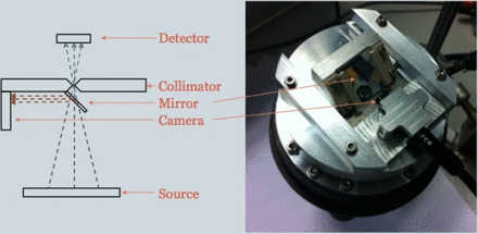 The compact hybrid optical-gamma imager. (Credit: Society of Nuclear Medicine and Molecular Imaging)The gamma camera comprising the scintigraphy aspect of the scanner camera detects radioactive signals emitted from the body after injection of a radionuclide, which interacts with specific physiological functions of the body. Nuclear medicine physicians can extrapolate information from the radionuclide’s activity.
Researchers evaluated the system in a clinical pilot study, which involved subjects undergoing routine molecular imaging procedures such as bone scans or imaging of the thyroid, eye or lymphatic system. Image resolution and acquisition time were optimized to less than five minutes by adopting a 1.5 mm-thick scintillator, which detects gamma rays as they are emitted from within the body, and a 1 mm pin-hole collimator, which functions as an aperture to narrow focus on a particular field of view.
The optical-gamma camera, which is still in development, was shown to be highly effective for imaging lymphatic and thyroid tissue, as well as drainage from the tear ducts, or lacrimal glands. Successful radionuclide absorption in these targeted areas was observed in tandem with optical images of surface anatomy.