3D-printed tissue featuring fluid channels mimics real organs for surgical training
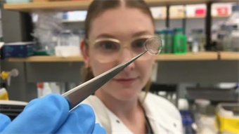
Marie Donlon | October 15, 2025Researchers at the University of Minnesota Twin Cities have developed a new 3D-printing method for creating realistic human tissue structures that could one day be used in the making of human models to train surgeons and doctors.
Currently, surgeons and doctors tend to rely on artificial models to practice delicate procedures. However, the majority of most of these training models meant to replicate human tissues tend to feel stiff, simple and unrealistic.
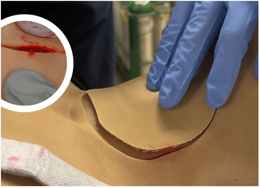 Source: The University of Minnesota Twin Cities
As such, there exists a gap between training models and real organs that has limited how well medical professionals can prepare before they eventually enter the operating room.
To develop models that look, feel and respond more like real human tissue, the Minnesota team developed a way to control the shape and size of microscopic patterns within the printed material that influences the strength and stretchiness of the tissues, thereby imbuing them with realistic mechanical properties.
Additionally, the team also created an accompanying mathematical formula to forecast how the tissues behave under stress.
Lending to the model’s life-like design was the addition of blood-like liquids during the printing process. The team noted that the microcapsules of the blood-like liquid are sealed so that the fluid would not evaporate or interfere with printing.
During trials of the realistic models, the new tissues scored higher on tactile feedback and cutting response than conventional replicas among surgeons
Going forward, the team plans to expand their work to mimic different organ shapes and functions.
An article detailing the work, “3D printed anisotropic tissue simulants with embedded fluid capsules for medical simulation and training,” appears in the journal Science Advances.