Enhanced Raman mapping of ferroelectric material properties
S. Himmelstein | July 06, 2023The analysis of dielectric response and other properties of functional crystals, such as perovskites, is challenging due to the inhomogeneous structures of such materials. An advanced microscopy technology engineered by researchers in Japan can facilitate analysis of the arrangement and fluctuation of electric polarization in ferroelectric materials.
The polarization-angle-resolved Raman microscope detailed in Communications Physics combines a half-wave plate with a Raman microscope to identify vibration states and directions in a material. The analytical system was used to examine a piezoelectric lead magnesium niobate-lead titanate crystal commonly incorporated in ultrasound equipment. These crystals have distinct dielectric and piezoelectric responses at their phase boundaries, and the titanium ratios responsible for their generation were effectively mapped with the new microscopy method.
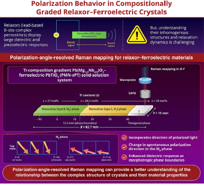 Polarization-angle-resolved Raman spectroscopy identified regions with enhanced charge accumulation in lead-based perovskites. Source: Ritsumeikan University
The measured spectra depicted a slower relaxation of polarization at certain phase boundaries; a slow response was demonstrated to enable the material to store a large amount of charge, leading to an enhanced dielectric response.
According to the researchers from Shimane University, Ritsumeikan University and the National Institutes for Quantum Science and Technology, this polarization-angle-resolved microscope could be used to optimize materials and their dielectric performance and improve ultrasound detection.