Watch: Imaging the internal structure of mini-brains
S. Himmelstein | January 27, 2021New imaging and labeling techniques enable visual assessment of the internal structure of brain spheroids and observation of the morphology of single neurons in 3D. Brain spheroids, termed mini brains by the international research team, are a cluster of different types of brain cells cultured from induced pluripotent stem cells.
Personalized mini-brains can be grown from stem cells generated from a sample of human hair or skin and could shed light on how brain disease progresses in an individual and how a patient may respond to drugs. Used as a research tool, including in drug development, mini brains will be more useful to researchers if 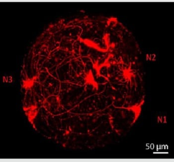 A 3D mini-brain imaged with a confocal microscope to reveal the structure of individual neurons. Source: Subashika Govindan et al.their structure can be accurately assessed without having to slice them for microscopy. This process makes it difficult to accurately determine internal structure or observe the structure of individual neurons. The sectioning process can also damage the spheroids, making subsequent imaging less valuable and informative.
Imaging techniques developed by researchers from University of Applied Sciences and Arts Western Switzerland, ARIMA Lifesciences PVT Ltd (India), Wyss Center for Bio and Neuroengineering (Switzerland) and Swiss Center for Applied Human Toxicology allow examination of the inside of intact spheroids. A novel technique for labeling individual neurons was combined with a method to render the sample completely transparent. A state-of-the-art custom module, including a bespoke sample holder and sensitive imaging detectors, was engineered to capture 3D images of entire intact mini-brains without slicing them. The 3D morphology of specific neurons and their anatomical distribution inside the mini-brains can now be visualized and analyzed.
The new approach could also enable imaging of large numbers of mini-brains, making it suitable for high-throughput screening for drug discovery or toxicity testing and reducing the need for animal testing.
A paper on the research is published in Frontiers in Bioengineering and Biotechnology.