Gel Electrophoresis Equipment: Process and Function
Engineering360 News Desk | July 07, 2015Gel electrophoresis equipment, instruments, and supplies separate nucleic acids or proteins on the basis of size, electric charge and other physical properties.
Gel electrophoresis is useful in forensics, biochemistry, genetics, microbiology and other applications requiring analysis of nucleic acid and protein molecule size and characteristics. Protein electrophoresis is also a common method for analyzing blood plasma in medical applications. Electrophoresis often occurs as a preparatory technique prior to cloning, DNA sequencing, Southern blotting, restriction fragment length polymorphism (RFLP), and analysis via mass spectrometry.
The Gel Electrophoresis Process Explained
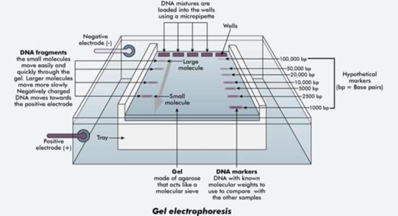 A typical gel electrophoresis apparatus. Image credit: Western Australia Department of Training and Workforce DevelopmentBefore electrophoresis begins, a user prepares and cools a liquid gel, pours it into a tray, and fits it with a toothed comb to create separations for each individual sample. A buffer fluid containing conductive ions is then added to the solidified gel in order to sustain current flow and maintain constant pH. The user injects a dyed nucleic acid or protein sample into the gel, and a power supply passes an electric current through it. Larger and longer molecules move slowly between the negative and positive supply poles, while smaller, shorter molecules move more quickly. Differently sized molecules therefore form distinct bands in the gel, which are analyzed or compared to other acid chains.
The gel functions as both an anticonvective and sieving medium in the electrophoresis process. As an anticonvective substance it suppresses heat buildup caused by current flow. As a sieve the gel retards the movement of molecules and retains the finished separation due to its matrix structure.
The video provides a basic overview of the gel electrophoresis process as described above.
Denaturing vs. Native
 The effect of SDS on a protein chain. Image credit: Davidson CollegeAn electrophoresis gel may be denaturing or native depending on the intended analysis. Denaturing disrupts the natural structure of the analyte and causes acids and proteins to unfold into simple chains by adding chemicals to the buffer solution. Typical denaturing additives are urea for nucleic acids and sodium dodecyl sulfate (SDS) for proteins.
Native electrophoresis maintains the analyte's natural structure during separation so that more complex biomolecular analysis can take place. The molecular complexes remain clustered or folded as they exist in the sample. This process is often more complex and unpredictable due to the difficulty in determining the effect of a molecule's shape and size on its mobility.
Dimensions
Electrophoresis may be one- or two-dimensional. One-dimensional electrophoresis involves one separation as described above, while the two-dimensional process adds a second separation to classify molecules by another parameter. Molecule separation properties include mass, isoelectric point, or complex mass in native state. Because molecules are unlikely to be similar regarding two different properties, 2-D electrophoresis is usually a more effective separation technique when compared to 1-D.
Types of Electrophoresis Processes
 Electrophoresis types, classified by gel type, separation type, or denaturing detergent. There are several specific electrophoresis process types, classified by gel type, separation type, or denaturing detergent type.
Polyacrylamide gel electrophoresis (PAGE) is one of the most common methods for separation of both nucleic acids and proteins, most often by mass. As its name implies, PAGE uses a polyacrylamide gel that is ideal for protein separation but also provides high resolving power for small DNA fragments.
SDS-PAGE is a common protein electrophoresis process that uses SDS as a denaturing agent. Its applications include determining molecular weight, identifying proteins, determining sample purity, identifying the existence of disulfide bonds, and detection or estimation of genetic diversity within a sample.
Isoelectric focusing (IEF) separates molecules by electric charge and is often paired with SDS-PAGE in a 2-D process involving separation by isoelectric point followed by mass. In the IEF process an ampholyte is added to an immobilized pH gradient (IPG) acrylamide gel. Proteins within the IPG gel will migrate to pH bands that correspond to their isoelectric point. Two-dimensional IEF/SDS-PAGE is used to analyze complex protein mixtures or tissue behaviors, partially characterize proteins, compare different physiological states of the same tissue, and purify proteins for further analysis.
Equipment Types
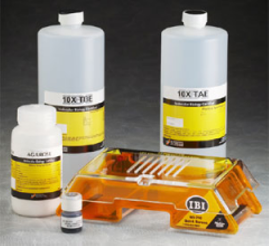
Complete electrophoresis sets or kits include most supplies necessary to the process. These may include a combination of gel trays, dyes, sample kits, and analysis materials. Some completely automated systems include a power supply and analysis software so that the user only needs to provide the analyte samples.
Process type, such as SDS-PAGE and IEF, and orientation are two common methods for describing systems. While PAGE and IEF are similar processes, they use different equipment and accessories. For example, IEF systems may include the required ampholyte solution or a specialized preparation device for the gel or samples.
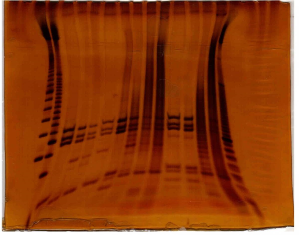 Smiling gel. System orientation is either vertical or horizontal. Horizontal (also known as flatbed) trays are rectangular and are ideal for analysis of large-volume samples. Vertical systems employ columnar gels and trays for analyzing various small sample quantities. Horizontal systems are sometimes considered advantageous due to their ability to evenly distribute heat over the entire gel surface. Uneven heat distribution can cause warped molecular bands, resulting in "smiling" effects as shown in the illustration.
Electrophoresis components
Electrophoresis components are often sold and procured separately. Common equipment includes dyes, trays, power supplies, electrodes, cables, gel mixtures, gel dryers, and chemicals such as denaturing agents, gel hardeners, and ampholytes.
Selection of an appropriate gel is most important to the electrophoresis process. Gels are layered into an upper stacking gel and lower separating/resolving gel. The stacking layer is low in acrylamide and has low pH, while the separating layer has a higher acrylamide concentration and higher pH. The differences between the two layers result in high resolution and more distinct bands for easier measurement.
Types of Gel
Three types of gel are used in the electrophoresis process:
- Agarose gels have low resolution but a high range of separation. They are employed in DNA fragment analysis.
- Polyacrylamide gels are used for protein and small-fragment DNA analysis.
- Starch gels are formed from partially hydrolyzed potato starch. They are slightly more opaque than agarose or polyacrylamide types and are used for protein electrophoresis.