Netflix algorithm used for faster biological imaging
Amy J. Born | April 01, 2019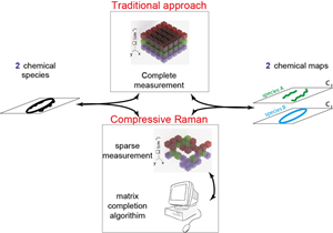 With the new compressive Raman approach, the researchers could acquire less spectral data than traditionally required and then use the matrix completion algorithm to fill in information not recorded. Source: Hilton De Aguiar, École Normale SupérieureAn algorithm originally created for a 2009 Netflix competition is shortening the time it takes to acquire Raman spectroscopy images of biological tissues.
The algorithm was intended to predict movie preferences of subscribers, but is finding a new use. The increased speed of this computational imaging approach could make the technique practical for clinical applications such as tumor detection and tissue analysis, the researchers reported in Optica.
Raman spectroscopy, named after physicist Sir C.V. Raman, is an imaging technique commonly used to identify unknown substances, identify polymorphs, track changes in molecular structures, evaluate residual stress on a crystal structure and assess the direction of orientation of molecules.
Among advantages for Raman spectroscopy are that it is non-invasive and it requires no sample preparation. One disadvantage for diagnostic purposes, however, is the amount of time it takes to gather images and process the data.
The researchers were able to speed up the process of biological imaging by acquiring only some of the data needed for Raman spectroscopy and using the algorithm entered in the Netflix contest to fill in the rest. They incorporated a spatial light modulator, which is a fast digital micromirror device, to replace slow cameras to speed up the imaging process.
“A very fast spatial light modulator made it possible to acquire images and skip data bits very quickly,” said Hilton de Aguiar, leader of the research team at École Normale Supérieure in France. “The spatial light modulator we used is orders of magnitude less expensive and faster than other options on the market, making the overall optical setup cheap and fast.”
Using a Raman microscope, the team demonstrated the ability to quickly capture images from brain tissue and single cells, which have high chemical complexity. The high level of data compression reduced the data up to 64 times.
The team plans to test this method with additional tissue types and will continue to work on reducing the scanning speed, with a goal of reaching sub-second image acquisition.