Microfluidic diagnostic sizes up cancer cells
S. Himmelstein | February 28, 2019Blood tests for detecting circulating cancer cells shed from a primary tumor into the bloodstream are being 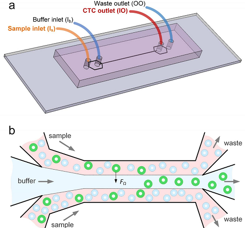 a) Configuration of the inlets and outlets. b) Diagram depicts how the microfluidics device separates cancer cells (green circles) from blood. Source: Ian Papautsky, University of Illinois Chicagoadvanced to speed diagnosis and eliminate the need for tissue biopsies. Biomedical researchers are challenged to design a device that can detect these single cells, often only present in extraordinarily small quantities.
Progress is reported in Microsystems & Nanoengineering by researchers from the University of Illinois Chicago and Queensland University of Technology, Australia, in the development of an inexpensive microfluidic tool that detects floating cancer cells in a blood sample. While other devices rely on affinity separation to induce biomarkers to latch onto tumor cells, the new system exploits size-dependent inertial migration to filter out circulating tumor cells based on cancer cell size relative to other blood sample constituents.
Previously-developed microfluidics devices designed to separate circulating tumor cells from blood have recovery rates between 50% and 80%. When applied to 5 ml blood samples spiked with 10 small-cell lung cancer cells, the new device returned a 93% detection rate. The method identified circulating tumor cells in six of eight samples from non-small lung cancer patients.
The team is preparing to conduct additional trials to verify the efficacy of this protocol and to enhance its accuracy with the addition of cancer cell DNA identification functionality.