Surgical Success with a 3D-Printed Skull
Engineering360 News Desk | April 13, 2016When a 15-year old patient was diagnosed with a tumor lodged in his sinuses, doctors at University of Michigan’s C.S. Mott Children’s Hospital knew that the effectiveness of minimally invasive surgery couldn’t be determined with traditional visualization approaches. The tumor, known as juvenile nasopharyngeal angiofibroma, is a mass that grows in the back of the nasal cavity and predominantly affects young male teens.
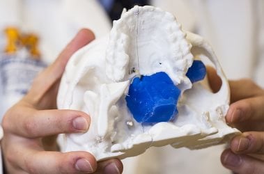 A 3D model guided surgery on a patient’s skull. Image source: University of Michigan Health Lab.To help determine optimal treatment, hospital researchers 3D-printed a replica of the patient’s skull. The polylactic acid model helped simulate the surgical procedure and allowed surgeons to plan the operation so as to avoid damaging nearby nerves and to minimize blood loss. Through preoperative embolization, the blood supply to the tumor was blocked off the day before surgery to decrease blood loss. A large portion of the tumor was then detached endoscopically and removed through the mouth. The remaining mass under the brain was taken out through the nose.
Doctors took pictures of the patient’s anatomy during the surgery and, later, compared it with pictures from the model. They were nearly identical.
Although medical application of the technology continues to gain attention, it isn’t entirely new. University of Michigan medical teams have used 3D printing for almost five years. Groundbreaking 3D printed splints made at the university have been used to treat babies with tracheobronchomalacia, which causes the windpipe to periodically collapse and prevents normal breathing. Doctors have also used 3D printing on a fetus to plan for a potentially complicated birth.