Carbon fiber in the biomedical industry
Chaithanya Ratakonda, M.D. | February 02, 2021Carbon fiber (CF) has numerous diverse applications across various technological fields, based on its unique physical, chemical and biological properties. CF is incredibly lightweight and has massive strength and chemical stability. Extensive studies have been conducted regarding carbon fiber applications to biomaterials, such as creating equipment structures to hold limbs during an X-ray.
Carbon, being relatively inert and a typical constituent of tissues, has several advantages over other materials. Since a large portion of living tissue is composed of carbon, it is readily accepted by the tissues. Over the last few decades, various carbon materials have been studied for their application in medical science.
With technological advancements, carbon fiber is now being controlled at the nanolevel to develop new composite materials that were not previously possible. Carbon nanofibers with the diameter in nanoscale can extensively enhance existing biomaterials. The unique nanofibers have created endless possibilities for improved applications in oncological treatment and regenerative medical science. With the first three-dimensional development of carbon nanofibers, it has been deduced that these materials can be used to provide exceptional scaffolding for bone tissue regeneration. The nanofiber material can be used as a scaffold in tendon and ligament repair and in the restoration of the hernial ring
In vitro biocompatibility evaluation of CFs
The process of constructing three-dimensional tissue through cell combination and scaffolds is called tissue engineering. The most essential aspect of this process is the adhesion and growth of the cells on the scaffold material. The biocompatibility of carbon fibers in tissue engineering has been researched extensively to provide more than one conclusion. Although some investigators state that CFs induce tissue growth, others do not. The cellular response to CFs actually depends on the degree of crystallinity of the material; therefore, not all CFs are biocompatible. The mixed opinions regarding the biocompatibility of CFs arise due to the use of fibrous carbon materials with different physical or chemical properties.
Fibroblasts are cells located in connecting tissues that secrete extracellular matrix (ECM). Due to their growth accelerating properties, fibroblasts are used in the regeneration of new tissues. An example of a typical cell culture model is the primary mouse embryonic fibroblasts (p-MEFs). Compared to other models, they are easier to maintain and proliferate a large number of cell production. However, fibroblasts lack certain surface molecules making them immunologically inert.
 Source: Adobe Stock
Methodology and requirements
Cell culture tests have been performed in several institutions across the globe. In a recent in vitro test conducted in the Indian Veterinary Research Institute in India, CF mesh was autoclaved. The cells obtained in the process were then washed thoroughly with Dulbecco’s Modified Eagle Medium (DMEM). The next day, the dead cells were removed, and the spindle-shaped fibroblast cells were collected. A well-executed process was carried out to get a single cell suspension. The CF mesh was then rewashed with DMEM and seeded in a humidified CO2 atmosphere. The seeded mesh was finally fixed for SEM examination.
Cultured p-MEF cells were observed under a phase-contrast microscope to assess the proliferation of cells. The monolayer of proliferated cells was completed in vitro and showed a characteristic growth pattern. The cultured in vitro cells clearly showed the morphology p-MEFs. An SEM examination evaluated the materials’ adhesion, proliferation and morphology.
The morphological examination disclosed that CF filaments were closely woven. The cells seeded on the walls of the CF mesh were spread uniformly as well. These seeded cells covered all of the CF mesh surfaces within a few days, indicating favorable biocompatibility of carbon fibers. The primary assumption that carbon being a typical constituent of tissues would not have a detrimental effect on them, encouraged scientists to experiment with carbon fiber implantations. Applications of carbon materials in medical science and research are becoming increasingly useful as they help in tissue regeneration and cure various human diseases.
Subcutaneous implantation of CFs
Four Wistar rats were anesthetized using a xylazine-ketamine anesthetic combination before subcutaneously implanting CF mesh on the spine of the animals. The animals underwent aseptic surgery to carry out the implants. The CF and mesh were retrieved a month after the surgery and were preserved in 10% formalin saline solution. The tissues were then processed by a routine paraffin embedding technique, and several sections were cut. These sections were studied once they had been stained with eosin (H & E) and hematoxylin stain.
Inferences
The retrieved sample displayed that CFs and mesh were covered with dense fibrous connective tissue. There were also no signs of infection or pus formation near the implanted biomaterials. Further SEM examination of the implanted material showed an excellent tissue response, with the material being surrounded with granular tissue. The implant clearly showed high biocompatibility of carbon fibers, as observed in high magnification.
Also, a histopathological examination showed multinucleated giant cells accompanied by eosinophils and mast cells. The muscle tissue surrounding the implant had a stable structure. Further, the increased number of fibroblasts revealed extensive fibrocellular reactions.
Carbon fibers in most cases did not have any detrimental effect on the foreign body. Instead, the tissues developed on carbon scaffold were extremely identical to the replaced tissue in both structure and functionality. The studies thus suggest that highly crystalline carbon fibers are not suitable for medical purposes. In contrast, amorphous carbon fibers are generally excellent biomaterials.
Carbon fibers in repair and reconstruction of abdominal wall defects
Reconstruction or repair of abdominal wall defects is very challenging. Surgeons have been using biomaterials like small intestine mucosa, expanded polytetrafluoroethylene, polyester fabric and human dura mater to reconstruct these wall defects. However, each of these biomaterials has its own set of problems resulting in a reduction of prevalent clinical acceptance. In contrast to all other biomaterials, carbon fibers provide immense structural strength along with a scaffold on which the repair takes place.
Conventional techniques that do not provide any structural reinforcement to the abdominal wall include the hazard of recurrence. To repair large abdominal wall defects, surgeons use prosthetic materials of different composition and properties to swap the missing tissue. Several tendon reconstructions have been successfully carried out with the use of carbon fibers as a biomaterial. CFs have numerous advantages over other similar materials, the most important being the inertness of carbon.
Carbon fibers in the repair of the gaps in flexor tendons
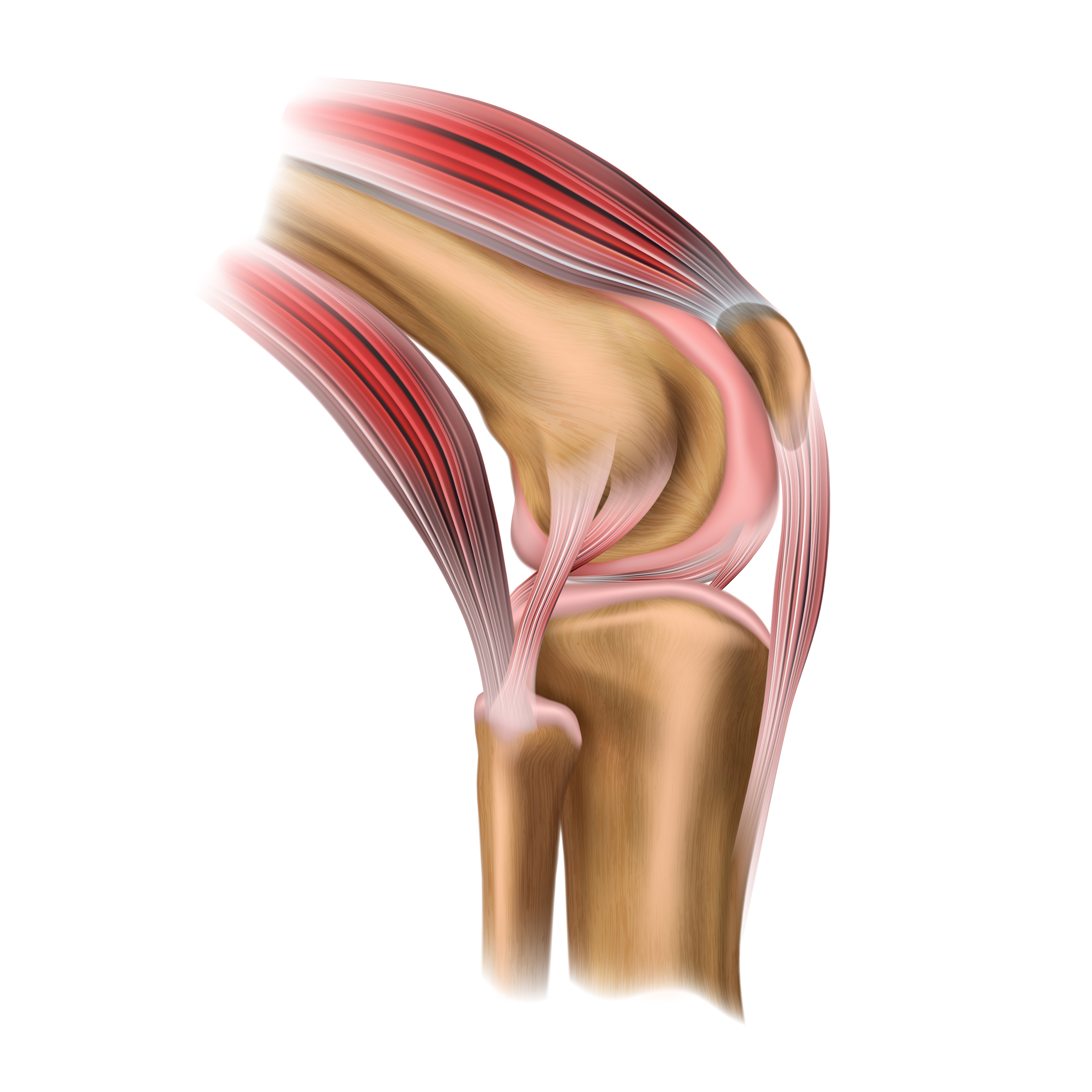
After a successful tendon reconstruction, certain gaps tend to form between cut ends. These gaps generate significant issues for the entire repairing process. Generally, carbon fibers are used to fill these gaps in animals like horses, sheep, rabbits and donkeys. The biocompatibility and non-carcinogenicity of CFs make it an excellent synthetic substitute. Carbon being an important constituent in animal tissue, it lets CF material smoothly integrate with the rest of the body without having any detrimental effects. The present medical study recommends the use of carbon fiber materials to repair the gaps in flexor tendons.
 Source: Adobe Stock
Clinical applications of carbon fibers
There have been many arguments regarding carbon fiber as a suitable biomaterial. The mechanism of removal of implanted CFs also has several associated controversies. Most medical studies have reported that CFs do not hinder tissue growth and act only as a scaffold for tissue proliferation. Since the physical, chemical and structural properties of carbon fibers depend on their microstructure, the various results obtained from evaluating different CFs as biomaterial can be explained with the help of these properties.
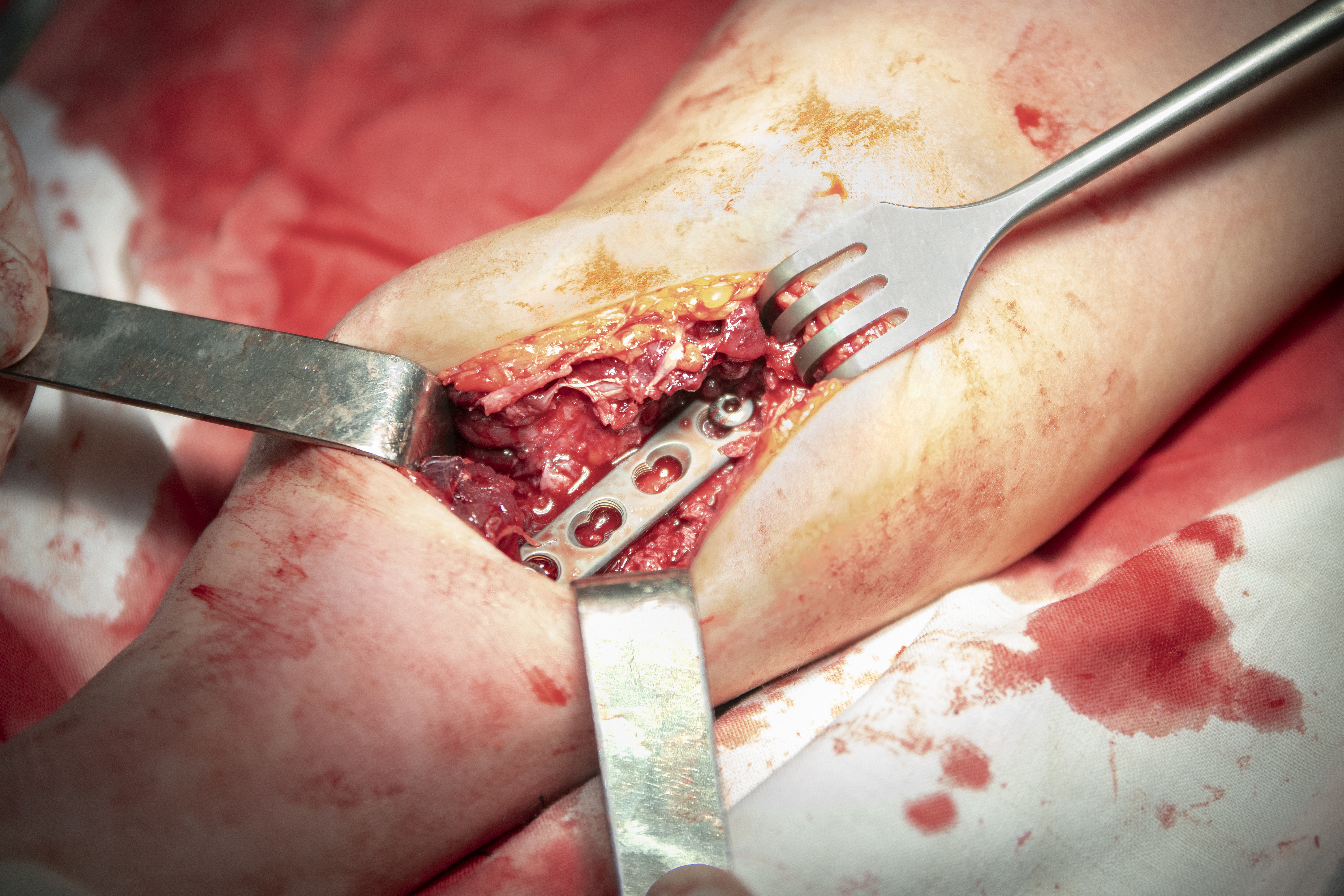
Although both sets of arguments provide logical explanations, thorough studies have shown that CF is indeed a better alternative to many other commonly used biomaterials. It is chemically stable, structurally strong and easily integrates with a living body with no long-term side effects. These properties enable carbon fiber to have numerous modern-day clinical applications like tendon and ligament repair and restoration of the hernial ring.
 Source: Adobe Stock
About the author
Dr. Ratakonda received her medical degree from NTR University of Health Sciences of Vijaywada, India. She is an award-winning medical writer and has more than 15 years of medical research experience, as well as experience in patient care. She specializes in the development of scientific and educational content, and describes herself as motivated by new challenges in the medical field.