Organ-On-a-Chip Device Models Heart Disease
S. Himmelstein | January 02, 2018Organ-on-a-chip devices offer an alternative to culturing cells on Petri dishes or experiments with lab animals to study diseases or new drug regimens. These systems mimic the functions of human organs, serving as potentially cheaper and more effective tools.
A new device for modeling atherosclerosis -- the constriction of blood vessels responsible for heart attacks and strokes -- was designed at Nanyang Technological University in Singapore. The organ-on-a-chip be used to study important inflammatory responses in cells that line the vessel in ways that could not be done in animal models. The researchers say it can also improve blood testing for patients.
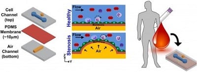 Atherosclerosis-on-a-chip microdevice for modeling stenosis and assessing blood health. Credit: Han Wei HouThe device fits on a single square-inch chip, consisting of two stacked chambers separated by a thin and flexible polymer membrane. The bottom contains air while the top contains a flowing fluid similar in mechanical properties to blood. Inside the fluid-filled chamber on top of the membrane, the researchers grow endothelial cells -- the cells that line the inside of blood vessels. The researchers pump air into the bottom chamber, so the membrane stretches like a balloon and forms a bubble that blocks the fluid flow. This process simulates the narrowing of a vessel.
The fluid-filled chamber constricts, causing the fluid to flow faster in some regions and slower in others. When the researchers grew the cells under continuous but slow fluid flow, endothelial cells were able to grow and express a protein called ICAM-1, which is associated with inflammation and is important in the development of atherosclerosis.
After replacing the cell culture media with human blood, more immune cells called monocytes bound to the endothelial cells in low-flow regions. Monocytes are mainly responsible for the accumulation of lipids, which eventually develop into the plaque that causes atherosclerosis.
These results on a chip are consistent with the widely accepted picture of the disease.
As a proof-of-concept experiment, the researchers pumped blood spiked with TNF-alpha, a protein indicative of inflammation, into their device. The inflamed blood caused more immune cells to bind to the endothelial cells than normal. Measuring the number of bound immune cells can reveal the level of inflammation in the blood, an indicator of early atherosclerosis. In contrast to other tests that just count the number of immune cells circulating in blood, this technique could more accurately assess early immune responses in patients.
The research is published in APL Bioengineering.