Higher Resolution Electrode Array Maps Brain During Surgery
S. Himmelstein | May 30, 2017Researchers from the University of California San Diego and Massachusetts General Hospital teamed up to redesign the electrode grid used by neurosurgeons to monitor neural activity and to stay clear of healthy tissue. The device provides higher resolution neural readings than existing tools used in the clinic and could enable doctors to perform safer, more precise brain surgeries.
Neurosurgeons use electrode grids to identify which areas of the brain are diseased in order to avoid damaging or removing healthy, functional tissue during operations. Despite their wide use, electrode grids have remained bulky and have not experienced any major advances over the last 20 years.
The new version is about a thousand times thinner — 6 micrometers versus several millimeters thick — than clinical electrode grids. This allows it to conform better to the intricately curved surface of the brain and obtain better readings. A much higher density of electrodes — spaced 25 times closer than those in clinical electrode grids — enables it to generate higher resolution recordings.
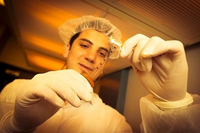 The PEDOT:PSS electrode grid is a new brain mapping device that can be used during brain surgery. (Credit: David Baillot/UC San Diego Jacobs School of Engineering)The researchers needed to shrink the size of the electrodes to pack them closer together, but miniaturizing metal electrodes increases their electrical resistance, resulting in more noisy readings. A conductive, thin, and flexible polymer called PEDOT:PSS was used instead of metal, resulting in the desired smaller electrodes without sacrificing electrochemical performance.
In side-by-side tests of the metal and polymer electrodes on four patients, the PEDOT:PSS electrode grid either performed similarly or slightly better than the standard electrode grid, recording with lower noise and higher resolution.
In one test, the team performed background readings of a patient’s brain waves both while the patient was awake and unconscious. The PEDOT:PSS electrode grid produced similar readings as the standard clinical electrode grid. In another test, the team monitored the brain activity of a patient undergoing epilepsy surgery. Both electrode grids identified normal functioning areas of the brain versus where the seizures were happening. The main difference is that the PEDOT:PSS electrode grid produced more detailed and higher resolution readings than the clinical electrode grid.
The next steps are to make higher density electrode grids for improved resolution and biocompatibility tests to see how long they can stay in the body before they experience biofouling.


