Coffee replaces staining agents in electron microscopy
Marie Donlon | January 12, 2026Researchers at the Institute of Electron Microscopy and Nanoanalysis (FELMI-ZFE) at Graz University of Technology (TU Graz) have discovered an environmentally friendly alternative to the staining agent used to ensure that the tissue structures of biological samples are visible under an electron microscope.
According to the research team, this alternative is ordinary espresso.
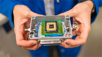
 Source: Claudia Mayrhofer — FELMI-ZFE
Currently, the standard staining agent used to make tissue visible under an electron microscope is uranyl acetate. Yet, some laboratories are not permitted to use this highly toxic and radioactive substance for safety reasons.
As such, the team sought an environmentally friendly alternative and the espresso-based stain used to treat samples in the lab proved as good quality as images of comparative samples prepared with uranyl acetate.
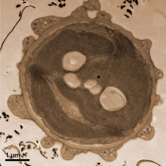
Inspired by the staining that occurs in coffee cups, the team demonstrated how the coffee stain could serve to make tissue visible under microscope by cutting the tissue samples into wafer-thin slices and fixing them onto sample holders. As a final step, the team stained the tissue before examining it under the electron microscope.
"Initial tests have shown that coffee stains biological samples and enhances contrasts," the team concluded. "Espresso provided comparatively very good contrast values, in some cases they were even better than with uranyl acetate.”
An article detailing the findings “Coffee — a ubiquitous substitute for uranyl acetate in staining of biological ultrathin sections for electron microscopy studies,” appears in the journal Methods.