Electrodes evolve inside living tissue
S. Himmelstein | April 17, 2023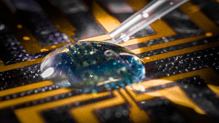 The injectable gel formulated to grow electrodes in living tissue is shown under test on a microfabricated circuit. Source: Thor Balkhed/Linköping University
A method for creating soft, substrate-free, electronically conductive materials in living tissue advances the merger of biology and technology to benefit medical and human-machine interface applications. Researchers in Sweden have demonstrated how injecting an enzyme-laden gel is altered by biological tissues to form an electrically conductive layer.
This achievement by scientists from Lund University, Linköping University and University of Gothenburg, who successfully grew electrodes in the tissue of zebrafish and medicinal leeches, paves the way for a new paradigm in bioelectronics. The surgically invasive implantation of rigid physical components to launch electronic processes in the body can be superseded by injection of a viscous gel.
The process entails use of a complex precursor system including an oxidase to generate hydrogen peroxide in situ, a peroxidase to catalyze oxidative polymerization, a water-soluble conjugated monomer, a polyelectrolyte with counterions for covalent cross linking, and a surfactant for stabilization. This formulation proved effective in inducing polymerization and subsequent gelation in different tissue environments to form electronically conducting material to specific biological substructures.
The approach could facilitate a variety of advanced medical systems, including pacemakers and brain-computer interfaces. The study published in the journal Science offers scope for the future fabrication of fully integrated electronic circuits in living organisms.


